Tobias Strickmann, PhD student at GEOMAR, is a jack-of-all-trades here on the Sonne. He collects environmental DNA samples for Véronique Merten, a postdoc at GEOMAR, and he also works with Anita Butterley, PhD student at the University of Tasmania, to filter water for proteomics samples. In addition to being a great team player, he also collects daily data for his own PhD research.
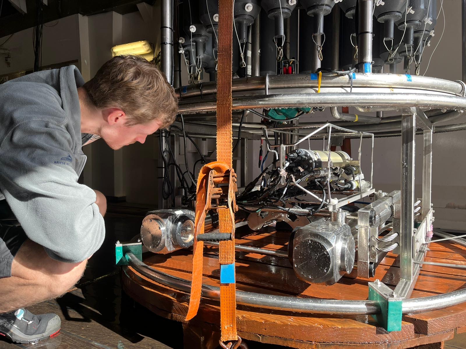
Like some scientists previously featured on this blog, he cares a lot about particles. However, instead of sampling using in-situ pumps, he uses cameras attached to a conductivity temperature depth (CTD) rosette. By photographing particles that pass the cameras’ fields of view as they move through the water column, he can determine the particle diameter and use that to calculate biovolume, which he in turn uses to estimate carbon content. By counting particles and calculating their carbon contents, he can estimate how the overall particulate carbon pool changes with depth, which, similar to Wan Zhang’s thorium activities, provides insight into the strength of the biological carbon pump (BCP). Combining particle count and size data from cameras with environmental parameters (like temperature and pH) from CTD instruments, he can investigate the effects of environmental conditions on the BCP.
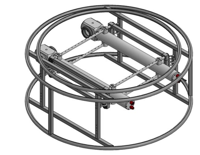
While many onboard the Sonne are partial to particles, perhaps the higher profile aspect of Tobias’s research is zooplankton portrait photography. He posts a “catch of the day” photo on the notice board—which those of us who study less charismatic subjects especially appreciate! These images come from the Plankton Imaging and Scanning Optics (PISCO) camera system, developed by engineer (and current shipmate) Anton Theileis and others from the Plankton Imaging research group— postdoc Veit Dausmann and group leader Jan Taucher. This bespoke camera has a special lens that allows the focus to shift while taking photos, resulting in crisp images. The amazing images come at a cost: Anton is on hand for constant tinkering, and the image volume is unwieldingly large. Contrast that to the Underwater Vision Profiler (UVP); although it has a fixed focus, it automatically crops the images it takes resulting in a more manageable data output.
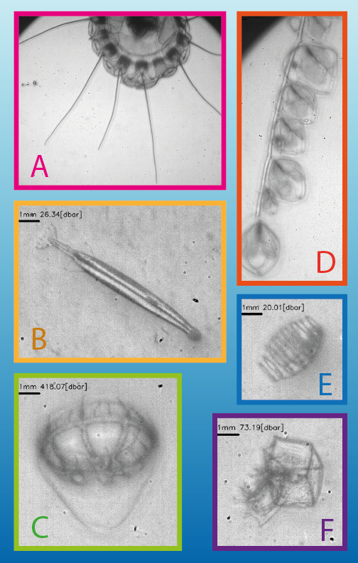
Tobias uses a custom classification system developed by Dausmann to identify the plankton in the photos, although he must first manually assign taxonomy to train the system on the dataset. As mentioned in our first blog post, the South Indian Ocean is understudied compared to other ocean regions, so Tobias is excited to contribute to the limited knowledge of zooplankton community composition in this area. An image is worth a thousand words, so tens of thousands of images will tell us volumes about particles and plankton!
By Charlotte Eckmann based on an interview with Tobias Strickmann.
A very good information from the sio-expedition. i hope to get more news from this exiting expedition.